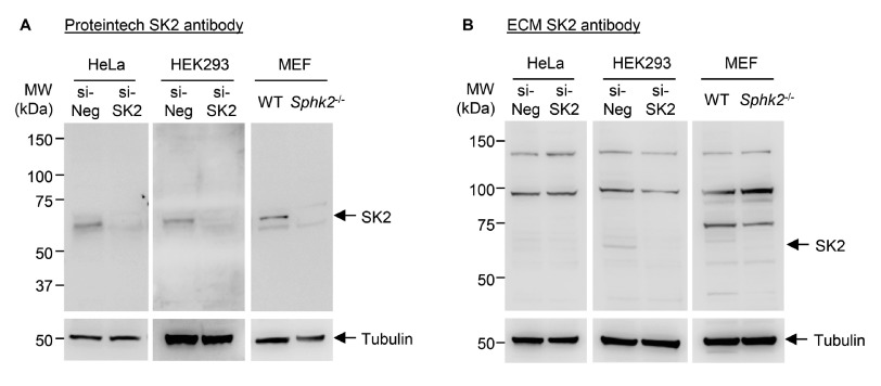
Figure 1. Immunoblot analyses of endogenous SK2 in multiple cell lines using two commercially available rabbit polyclonal anti-SK2 antibodies.
Immunoblot analyses of lysates from HEK293 and HeLa cells treated with scrambled control siRNA (si-Neg) or SK2 siRNA (si-SK2), and lysates from wildtype (WT) or Sphk2 -/- MEFs. An equal amount (40 µg) of total protein from each sample was run in duplicate. After transferring to nitrocellulose and blocking, the membrane was separated and duplicate samples were probed with either ( A) Proteintech rabbit anti-SK2 antibody or ( B) ECM Biosciences rabbit anti-SK2 antibody. SK2 membranes were imaged using a 4 min exposure. The expected band size for SK2 is ∼65 kDa. Membranes were re-probed with mouse anti-α-tubulin antibody as a loading control (2 min exposure), which was detected at 55 kDa as expected. Consistent results were observed from 2-3 (HEK293 and MEF) or 3-4 (HeLa) independent experiments for each antibody.

