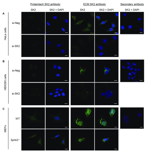
Figure 3. Immunofluorescence staining analyses of endogenous SK2 in multiple cell lines using two commercially available rabbit polyclonal anti-SK2 antibodies.
( A) HeLa or ( B) HEK293 cells were treated with scrambled control siRNA (si-Neg) or SK2 siRNA (si-SK2), and endogenous SK2 (green) was visualised by immunofluorescence staining and confocal microscopy, using Proteintech rabbit anti-SK2 antibody or ECM Biosciences rabbit anti-SK2 antibody. ( C) Wildtype (WT) or Sphk2 -/- MEFs were seeded, and endogenous SK2 (green) was visualised by immunofluorescence staining and confocal microscopy, using Proteintech rabbit anti-SK2 antibody or ECM Biosciences rabbit anti-SK2 antibody. Nuclei were stained with DAPI (blue). For each cell line, background staining was examined by staining cells (si-Neg or WT cells) with secondary antibody and DAPI only, and collecting images using both 488nm and 405nm lasers (SK2 + DAPI). Images were taken at 40× magnification; scale bars = 10 µm. Images shown are representative of more than 100 cells from each experiment, and these results were consistent over three independent experiments for each cell line.

