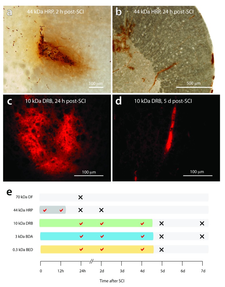
Figure 10. Temporal pattern of blood-spinal cord barrier disruption after SCI.
( a) Extravasation of HRP (44 kDa) extending radially out from sites of injury at 2 h post-injury. ( b) HRP is confined to the lumen of blood vessels at 24 h post-injury with no visible leakage around sites of injury. ( c) Extensive leakage of the 10 kDa dextran rhodamine B is observed around sites of injury at 24 h post-injury. ( d) at 5 days post-injury, 10 kDa dextran rhodamine B was always confined to the lumen of blood vessels. ( e) diagrammatic summary of the period of blood-spinal cord barrier disruption after SCI vs the molecular size of the permeability tracers. The tracers; dextran fluorescein, 70 kDa DF; horseradish peroxidase, HRP 44 kDa, dextran rhodamine B (DRB 10 kDa), biotin-dextran-amine (BDA 3 kDa), biotin-ethylene-diamine (BED 0.3 kDa) were injected systemically 20 minutes prior to collection of tissue. A tick mark indicates the presence of visible tracer extravasation whereas a cross indicates that the tracers were confined to the lumen of blood vessels.

