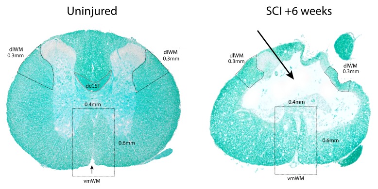
Figure 1. Transverse sections of thoracic (T10) level spinal cords stained with luxol fast blue (LFB).
Left is from an uninjured control animal and right is from the injury centre in an animal 6 weeks after SCI. The outlined regions (black dashed lines) mark the areas used for measurements of dorsolateral and ventromedial white matter (dlWM and vmWM respectively). The red outline indicates the position of the dorsal column corticospinal tract (dcCST) at the base of the dorsal column. The dlWM regions were defined as all stained white matter from the ventral side of the dorsal horn down to 0.3 mm along the outer circumference of the cord. Both the left and right dlWM areas were measured. The vmWM was defined as the total area of stained tissue within a rectangle measuring 0.4 mm × 0.6 mm centred on the sulcal fissure (small black arrow in left image). The absence of most of the dorsal column and dcCST at 6 weeks post-injury is indicated by the large arrow in the right image.

