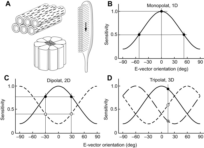
Fig. 2.
Structural and physiological properties of polarization-sensitive invertebrate photoreceptors. (A) Structural basis of polarization sensitivity in invertebrate photoreceptors. Right, microvilli form the rhabdomere of the photoreceptor. Light travels orthogonal to the microvilli (see arrow). Upper left, the visual pigment molecules with their elongated chromophores (black bars) reside in the microvillar membrane. Alignment of both chromophores and microvilli produces strong polarization sensitivity in the photoreceptor. Lower left, arthropod photoreceptors are grouped to form retinulae. (B–D) E-vector sensitivity functions. (B) A one-dimensional system (monopolat). (C) A two-dimensional system (dipolat) with 90 deg phase-shifted sensitivity functions. (D) A three-dimensional system (tripolat) with 60 deg phase-shifted sensitivity functions. In B–D, polarization sensitivity (PS)=5.0, degree of linear polarization (d)=1.0. A value of 0 deg indicates horizontal e-vector orientation and 90 deg indicates vertical e-vector orientation. Thin straight lines indicate values of selected data points (circular symbols) on the sensitivity and e-vector axes.

