Fig. 7.
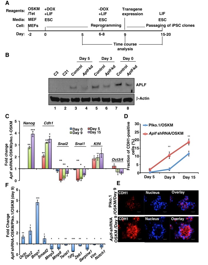
APLF regulates genes implicated in MET during the generation of iPSCs from fibroblasts. (A) Schematic representation for the time frame (not to scale) for the generation of iPSCs. Dox, doxycycline. (B) Western blot analysis for the expression of APLF in control and Aplf-kd MEFs at different stages of reprogramming. (C) qRT-PCR analysis was performed from samples collected at different days of reprogramming, and fold changes in gene expression were calculated in Aplf-kd cells relative to those in control cells. (D) CDH1-positive cells were determined by FACS analysis at different stages of reprogramming. (E) Immunofluorescence study of the expression of CDH1 in control and Aplf-kd MEFs at day 9. Nucleus, Hoechst 33258. (F) The same set of samples as described in E were analyzed for the expression of EMT-specific genes by performing qRT-PCR. Bar graphs represent the fold change in expression relative to in cells without shRNA. Error bars are s.e.m., n=3, *P<0.05, **P<0.01, ***P<0.001 (Student's t-test). Scale bar: 20 µm.

