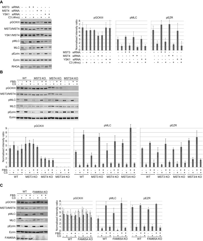
Fig. 3.
RHO and FAM65A do not regulate MST kinase activity. (A) The majority of GCKIII kinase activity in HeLa cells comes from MST4, with MST3 contributing to the remainder, both independently of RHO activity. HeLa cells were transfected with the indicated siRNA pools or non-targeting siRNA pool as control. 72 h post transfection, the cells were treated as indicated with TAT-C3 (C3) for 4 h, before lysis and analysis by immunoblotting with the indicated antibodies. P prefix indicates phosphorylated forms of the indicated proteins. Active phosphorylated GCKIII (pGCKIII) is resolved as a doublet, with the lower more-intense band corresponding to MST4, and the higher weaker band corresponding to MST3. TAT-C3 inactivated RHO, as manifested by a reduction in pMLC and pEzrin (pEZR) levels, but neither TAT-C3 nor YSK1 depletion affected pGCKIII levels. Bar graphs on the right-hand side of the blots display the quantifications of the indicated phosphorylated proteins. pGCKIII and pEzrin levels were normalised to total Ezrin levels as loading control, whereas pMLC was normalised to total MLC levels (arbitrary units). Quantification was performed on three independent experiments. Error bars=s.d. (B) GCKIII kinase activity comes from MST3 and MST4 and is not regulated by RHO. Wild-type (WT), MST3, MST4 or MST3 MST4 (MST3/4) double CRISPR knockout (KO) HeLa cells were starved for 24 h and treated as indicated with TAT-C3 for 4 h, before being stimulated by 10% FBS (15 min). The cells were then lysed and analysed by immunoblotting with the indicated antibodies. Stimulation activated RHO, as revealed by an increase in pMLC levels, whereas TAT-C3 inhibited RHO. Neither treatment affected pGCKIII levels, whereas double MST-knockout abrogated pGCKIII. Bar graphs below the blots display the quantifications of the indicated phosphorylated proteins. pGCKIII and pEzrin were normalised to total Ezrin levels as loading control, whereas pMLC was normalised to total MLC levels (arbitrary units). Quantification was performed on three independent experiments. Error bars=s.d. (C) GCKIII kinase activity is not regulated by FAM65A or RHO. WT or FAM65A CRISPR KO HeLa cell lines were starved for 24 h and treated as indicated with TAT-C3 for 4 h, before being stimulated by 10% FBS (15 min). The cells were then lysed and analysed by immunoblotting with the indicated antibodies. Neither FAM65A loss nor RHO activation–inactivation by FBS or TAT-C3 affected pGCKIII levels. Bar graphs on the right-hand side of the blots display the quantifications of the indicated phosphorylated proteins. pGCKIII was normalised to either total Ezrin (light grey bars) or total MST3 and MST4 (dark grey bars). pEzrin was normalised to total Ezrin, whereas pMLC was normalised to total MLC (arbitrary units). Quantification was performed on three independent experiments. Error bars=s.d.

