Fig. 1.

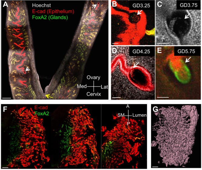
Confocal imaging of pregnant mouse uterus and cycling human endometrium. (A) Optical z slice showing two-thirds of both horns of a GD4.25 mouse uterus attached at the cervix (yellow arrow) stained with nuclear marker Hoechst (gray), epithelial marker E-CAD (red) and glandular marker FOXA2 (green). (B-D) Identification of blastocysts (white arrows) in optical slices of intact uteri at GD3.25 (B), GD3.75 (C) and GD4.25 (D). (E) Epiblast at GD5.75. (F,G) Three z slices (F) through a full-thickness segment of human endometrium stained for E-CAD (red) and FOXA2 (green), and corresponding surface rendering of the same specimen based on E-CAD staining (G). Scale bars: 500 μm in A,F,G; 50 μm in B-E. Lat, lateral; Med, medial; A, anterior; P, posterior; SM, smooth muscle.
