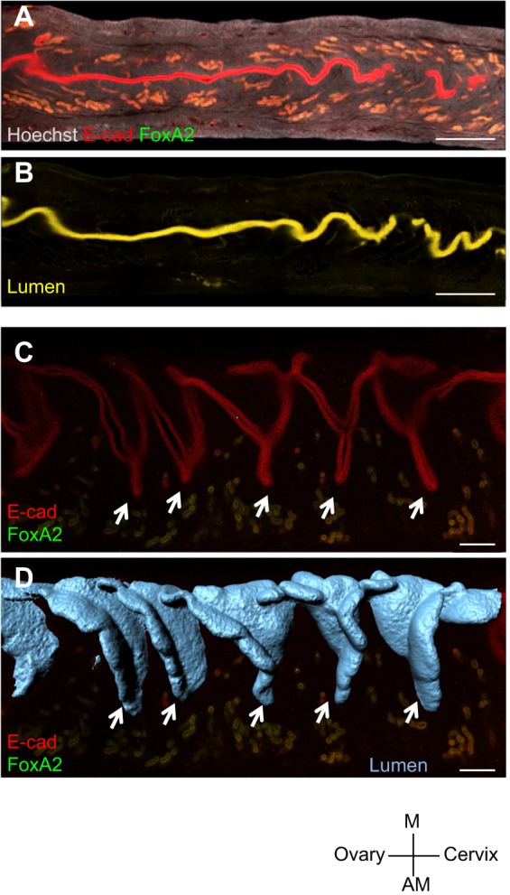
Fig. 2.

Uterine crypts are generated by folding the luminal epithelium. (A,B) Optical slices through a segment of the uterus at GD4.25 immunolabeled for FOXA2 signal (green) and ECAD signal (red) (A), and the resulting subtraction of FOXA2 from ECAD signal to obtain the uterine lumen (yellow) (B). (C) Optical slice showing uterine crypts (arrows) revealed by luminal epithelial staining at GD4.25. (D) Luminal folds (arrows) in the 3D surface model of the uterus coincide with crypts in the optical slice. Scale bars: 500 μm in A,B; 200 μm in C,D. M, mesometrial; AM, anti-mesometrial.
