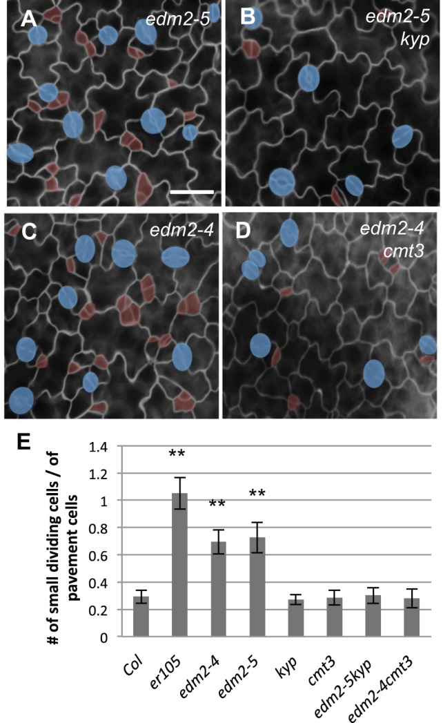
Fig. 7.

cmt3 and kyp mutations suppress the stomatal defects in edm2 mutants. (A-D) Epi-fluorescence microscope images show 3-dpg adaxial cotyledons of edm2-5 (A), edm2-5 kyp (B), edm2-4 (C) and edm2-4 cmt3 (D). Scale bar: 30 μm (in A, for A-D). cmt3 and kyp were epistatic to edm2 mutations. (E) Histogram displaying the ratio of the total number of small dividing cells over that of the pavement cells in different mutant backgrounds. *P<0.05, **P<0.01; one-way ANOVA with Bonferroni multiple comparison (n=6; ±s.d.).
