Fig. 4.
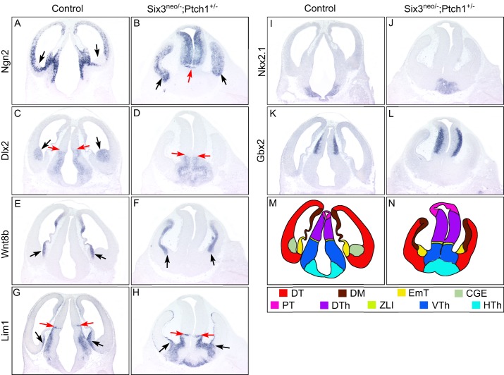
Elevated Shh signaling fails to rescue brain defects in Six3neo/– embryos. Coronal sections of E12.5 control and Six3neo/–;Ptch1+/– forebrains. In controls, the dorsal telencephalic marker Ngn2 is excluded from the caudal ganglionic eminence (CGE) (A,C, arrows), which is identified by Dlx2 expression (C). By contrast, Ngn2 is expressed throughout the telencephalic neural epithelium in Six3neo/–;Ptch1+/– embryos (B, arrows). Rostral-expanded diencephalon separates the telencephalic vesicles (B, red arrow). Wnt8b labels the dorsal midline (DM) of the telencephalon and the eminentia thalami (EmT) in control and mutant samples (E,F, arrows). The presence of the EmT is confirmed by the expression of Lim1 (G,H, black arrows). Lim1 is also a marker for the zona limitans intrathalamica (ZLI), the ventral thalamus (VTh) and the hypothalamus (HTh). Compared with that in wild-type embryos (G, red arrows), the region of Lim1 expression is slightly smaller and located more ventrally in Six3neo/–;Ptch1+/– embryos (H, red arrows). Similarly, Dlx2, another marker for the VTh and HTh, is expressed more ventrally and in a smaller area in the mutant embryos (D, red arrows) than in controls (C, red arrows). The presence of the HTh is marked by Nkx2.1 expression in control and mutant embryos (I,J). Ngn2 and Gbx2 are expressed in the dorsal thalamus (DTh), and their expression domain is enlarged in the mutant brain (B,L) compared with that in controls (A,K). (M,N) Summary of the expression analysis results. DT, dorsal telencephalon; PT, pretectum. n=3 control; n=3 Six3neo/−;Ptch1+/−.

