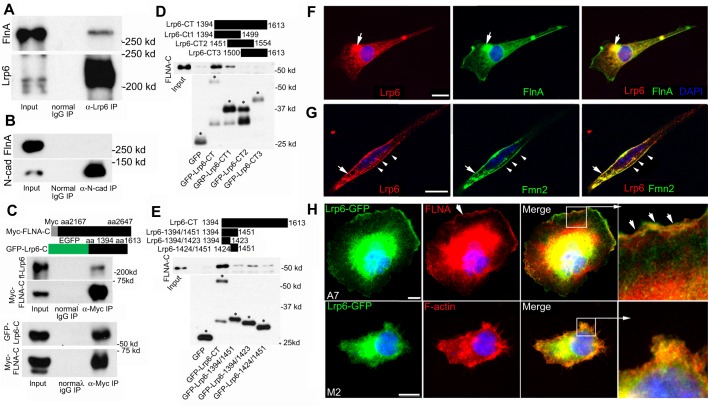
Fig. 5.
FlnA and Fmn2 interact and overlap with components of the Wnt signaling pathway. (A) Endogenous Lrp6 interacts with endogenous FlnA. Co-immunoprecipitation was performed by incubating E13.5 brain lysates with normal IgG and anti-Lrp6 antibody. (B) Control N-cadherin could not precipitate FlnA under the same condition as in A. (C) Conversely, myc-tagged FLNA C-terminus immunoprecipitated both full-length Lrp6 (upper panel) and its GFP-tagged C-terminus (lower panel). Schematics illustrate the myc-fused FLNA C-terminus and GFP-fused Lrp6 C-terminus fragments. (D) FLNA binding to Lrp6 is localized to a region of the C-terminus, as contained within Lrp6-CT1 (amino acids 1394-1499). The Lrp6 C-terminus (Lrp6-CT) was subdivided into three fragments (CT1, CT2 and CT3), each overlapping by ∼50 amino acids. The upper panel demonstrates that FLNA-C is pulled down by Lrp6-CT and Lrp6-CT1, and the lower panel shows the purified Lrp6 C-terminus fragments. Asterisks indicate bands of correct sizes for the fragments. (E) The Lrp6 binding site for FlnA was further refined to the Lrp6 C-terminus (amino acids 1394-1423). Regions of Lrp6 fragments are illustrated at the top. Western blot analysis shows that the strongest interaction with FlnA occurs with Lrp6-1394/1423, although weaker binding is also seen with Lrp6-1424/1451, suggesting that multiple site interactions exist for FlnA binding. Asterisks label correct sizes of undegraded fragments. (F) Immunocytochemistry shows overlapping Lrp6 and FlnA expression on the cell membrane and in cytoplasmic vesicles (arrows) of cultured neural progenitors. (G) Lrp6 also overlaps with Fmn2 on the cell membrane (arrow) and in perinuclear vesicles of cultured neural progenitors (arrowheads). (H) Lrp6 localization is dependent on FlnA. Loss of FLNA in M2 melanoma cells leads to diminished Lrp6 localization along the lamellapodia, as compared with A7 FLNA-repleted cells (arrowheads). Higher magnification images of the boxed regions are shown to the right. Nuclei are counterstained with DAPI (blue) in all images. Scale bars: 10 μm in F-H.

