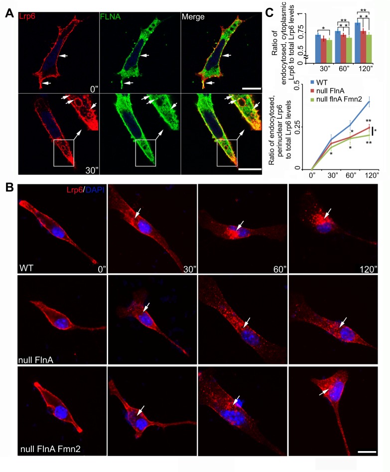
Fig. 6.
Loss of FlnA and Fmn2 impairs the rate of Lrp6 endocytosis. (A) Fluorescent photomicrographs show that FlnA (Fluorescein) and Lrp6 (Rhodamine) colocalize and undergo endocytosis into cytoplasmic vesicles. Neural progenitor cells transiently expressing Lrp6 were incubated with anti-Lrp6 antibody on ice for 40 min, then either fixed immediately (0 min) or incubated at 37°C for endocytosis (30-120 min). The top row shows that surface Lrp6 overlaps with FlnA on the cell membrane prior to endocytosis; the bottom row indicates that FlnA is localized with Lrp6-endocytosized vesicles (arrows) 30 min post-incubation. (B) Loss of both FlnA and Fmn2 impairs the rate of Lrp6 endocytosis, as compared with loss of FlnA alone. Fluorescent photomicrographs show representative Lrp6 endocytosis in WT, Flna null and Flna+Fmn2 null neural progenitors cultured in medium containing Lrp6 antibody and Wnt3a. Lrp6 endocytosis (arrows) was examined at various time points post-incubation at 37°C by staining for anti-Lrp6 with secondary antibody (Rhodamine). (C) Quantification of the rate of Lrp6 endocytosis into the cytosol and then to the perinuclear region. (Top) Lrp6 endocytosis in Flna+Fmn2 null cells is decreased by 9.7%, 11.3% and 17% at 30, 60 and 120 min, respectively, compared with that in WT cells. The rate of endocytosis was also significantly slower than that in Flna null cells, by 3.0%, 5.8% and 6.5% at these same time points. (Bottom) The rate of Lrp6 transport to the perinuclear region in Flna+Fmn2 null cells was decreased by 25%, 31% and 48% compared with that in WT cells, and by 14%, 3.2% and 17% compared with Flna null cells. Total or endocytosed Lrp6 levels were measured with ImageJ. *P<0.05, **P<0.01, two-tailed t-test. Error bars indicate s.d. Scale bars: 10 µm in A,B.

