Fig. 2.
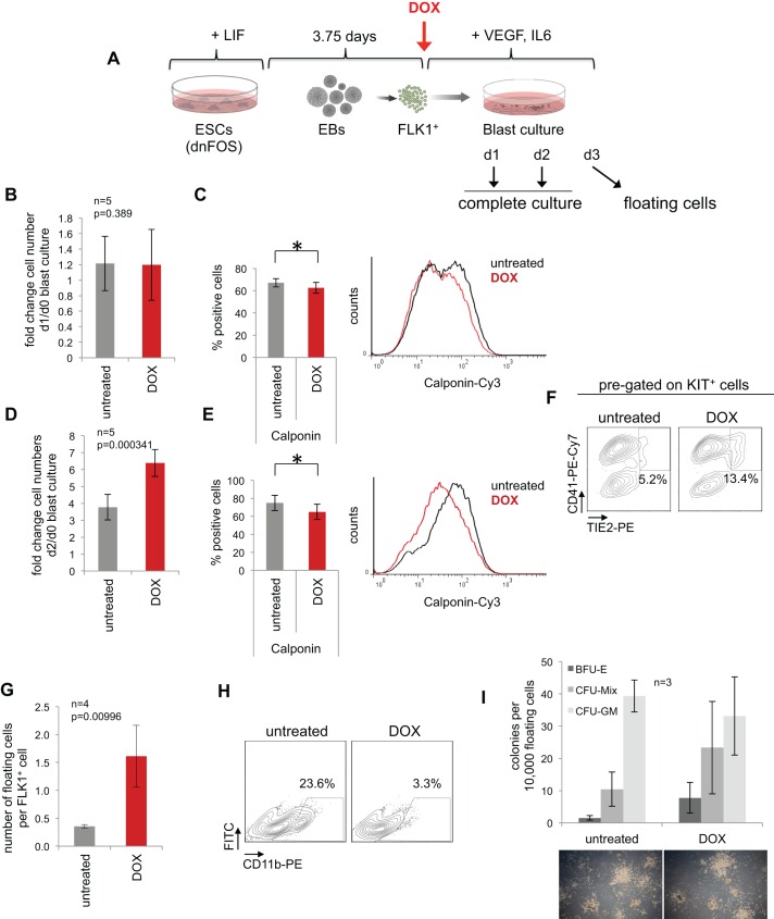
AP-1 inhibition at the hemangioblast stage enhances cell proliferation and shifts the balance between vascular and blood cell development. (A) Experimental setup: dnFOS ESCs were differentiated for 3.75 days as EBs. FLK1+ HB cells were purified and subsequently cultured±1 µg/ml DOX under blast culture conditions for 1 day, 2 days or 3 days before either complete cultures (day 1 and day 2) or floating cells (day 3) were analysed. (B) Fold changes of cell numbers at day 1 compared with day 0 (=seeded cell numbers) of complete dnFOS blast cultures with and without DOX induction (data are mean ±s.d., n=5, t-test). (C) Complete day 1 dnFOS blast cultures with and without DOX induction were assessed by intracellular SM cell-specific calponin staining (data are mean ±s.d., n=4, t-test, *P=0.019). A representative flow cytometric analysis and the summary of three experiments is shown (for other smooth muscle cell markers and statistical summary, see Fig. S2). (D) Fold changes of cell numbers at day 2 compared with day 0 (=seeded cell numbers) of complete dnFOS blast cultures with and without DOX induction (data are mean ±s.d., n=5, t-test). (E) Complete day 2 dnFOS blast cultures with and without DOX induction were assessed by intracellular SM cell-specific calponin staining. (data are mean ±s.d., n=3, t-test, *P=0.038). A representative flow cytometric analysis and the summary of three experiments is shown (for other smooth muscle cell markers and statistical summary, see Fig. S2). (F) The cell composition of complete day 2 dnFOS blast cultures ±DOX was analysed by flow cytometry using antibodies against KIT, TIE2 and CD41. A representative contour plot of pre-gated KIT+ cells is shown. (G) The number of dnFOS floating cells with and without DOX induction at day 3 per FLK1+ cell that was seeded at day 0 (data are mean±s.d., n=4). (H) Floating cells of day 3 dnFOS blast cultures with and without DOX induction were harvested and stained with a CD11b-specific antibody prior to flow cytometric analysis. Representative contour plots are shown. (I) Floating cells derived from day 3 dnFOS blast cultures with and without DOX induction were harvested and plated into methylcellulose medium for a hematopoietic colony assay (in the absence of DOX). After 10 days, colonies were counted and classified as BFU-E, CFU-Mix and CFU-GM (data are mean ±s.d., n=3). Examples of colonies are shown at the bottom.

