Fig. 5.
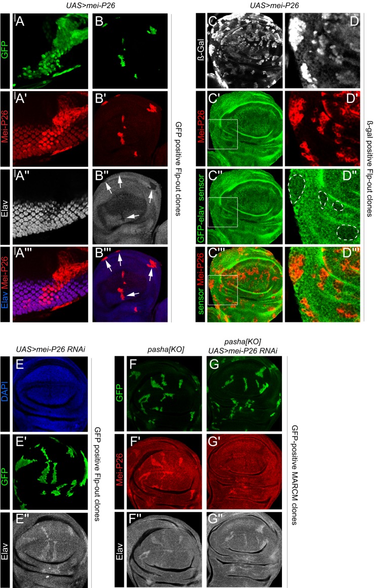
Mei-P26 represses elav via its 3′ UTR. Shown are (A) eye imaginal disc and (B-G) pouch regions of wing imaginal discs. (A-B‴) Clonal activation of Mei-P26 leads to cell-autonomous decrease in Elav protein in both a high-expression domain (photoreceptors, A) and a low-expression domain (wing pouch, B, arrows). (C-D‴) Ectopic Mei-P26 represses the tub-GFP-elav-3′ UTR sensor. Although the effect is quantitatively mild, it is more clearly observed in higher magnification clones (D, outlined regions). (E) Clonal knockdown of mei-P26 causes cell-autonomous increase in basal Elav. (F-G″) MARCM analysis of pasha[KO] clones. (F) Control pasha clones derepress both Mei-P26 and Elav proteins. (G) Knockdown of mei-P26 reverses the accumulation of Mei-P26 protein in pasha clones, but does not reliably superactivate Elav. At least five imaginal discs were assayed for each genotype, and each set of stainings was performed at least twice; representative clones are shown.

