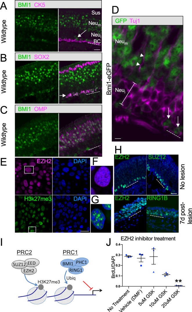
Fig. 6.

BMI1 and associated PRC proteins in vivo and in GBC cultures. An immunohistochemical panel applied to OE sections from 3-week-old wild-type mice confirms colocalization of BMI1 and markers for GBCs or mature neurons, whereas HBCs, immature neurons and sustentacular cells are BMI1−. (A) Mouse anti-BMI1-labeled basal cells (arrow) are not labeled by antibody to cytokeratin 5 (CK5) and are situated just above the CK5+ horizontal cell layer. OE cell layers are indicated as in Fig. 5. (B) SOX2 and rabbit anti-BMI1 colocalize extensively in basal cell layers (arrow); the apical SOX2+ sustentacular cell layer is not BMI1 labeled. (C) OMP+ neurons are BMI1+, whereas the immature neuron cell layers below the OMP+ neurons and above the GBCs do not express BMI1. (D) Tissue sections from Bmi1GFP knock-in mice confirm the BMI1 expression pattern. High-magnification view of the basal region of the OE. Tuj1 antibody labels immature differentiating neuron somata below the OMP+ layers (bracket). Note that the Tuj1-labeled cells do not express GFP. Arrows mark GFP+ basal cells; GFP is also present in the mature neuronal layers apical to the Tuj1+ immature cells; arrowheads mark a Tuj1+ dendrite. Dashed line marks basal lamina. (E-J) Additional PRC1/PRC2 proteins were localized to nuclei in GBC cultures (E-G) and in vivo (H). The H3K27me3 mark labels cultured GBCs (E), consistent with PRC2 transcriptional repression (see schematic I). Perturbation of Bmi1 in GBC cultures resulted in gene expression changes but no obvious phenotype at 48 h (see Fig. 4D). However, treatment with GSK343, an inhibitor of the PRC2 component EZH2, resulted in a rapid decrease in proliferation (J). **P=0.0037, ANOVA, versus vehicle (dimethyl formamide). Error bars indicate s.d. Scale bars: 25 μm A-C,E,H; 5 μm in D.
