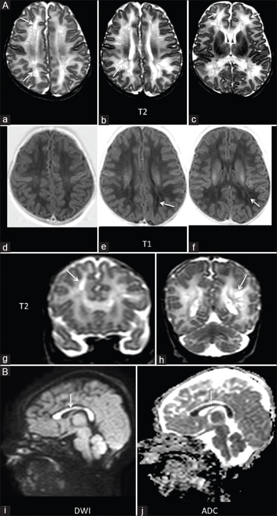
Figure 2 (A and B).

(A) A 20-day-old term neonate with history of asphyxia and two episodes of left-sided focal seizures. Multiple small areas of abnormal signal intensity appearing hyperintense on T2 and isointense on T1WI seen in the periventricular white matter bilaterally, predominantly in the frontal lobes (arrow, a-h). (B) Restricted diffusion was noted in the entire corpus callosum (arrow, I and j)
