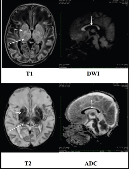
Figure 3.

A 13-day-old, term neonate with respiratory distress, hypotonia, and seizures. MRI revealed abnormal signal intensity in the thalami and the basal ganglia, appearing hyperintense on T1WI and hypointense on T2WI images (long and short arrow). The entire corpus callosum showed restricted diffusion. (White arrow)
