
Figure 5 (A and B).
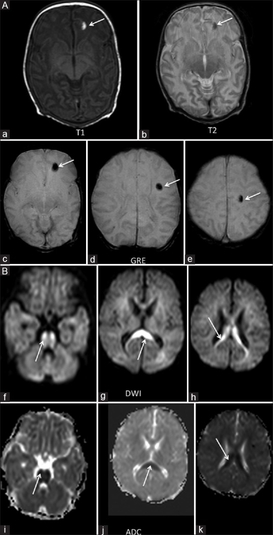
(A) An 11-day-old term neonate with history of seizures. Three focal areas of blooming were seen in the left frontal lobe on GRE images (arrow c-e). These areas appeared hypointense on T2WI and hyperintense on T1WI (arrow a and b), suggestive of a subacute hemorrhage. (B) The neonate also had areas of restricted diffusion in the corpus callosum involving the body (arrow h and k), splenium (arrow g and h) and the pons (arrow f and i)