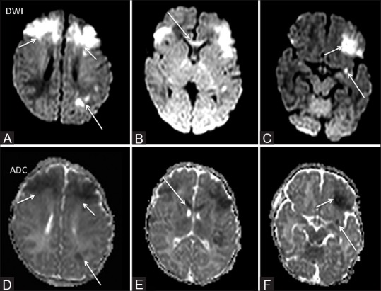
Figure 6 (A-F).

A 5-day-old term neonate a suspected case of HIE. Restricted diffusion was noted bilaterally in the high frontal region (short arrow A and D), left basifrontal region (short arrow F and C) and genu of corpus callosum (arrows B and E). Small focus of restricted diffusion was also noted in the left perisylvian region (long arrow F and C) and in the left parietal lobe (long arrow A and D)
