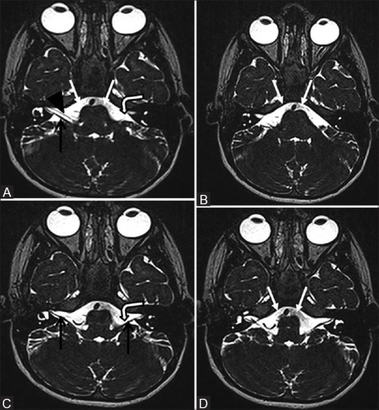
Figure 2 (A-D).

Four (A–D) contiguous 0.7-mm thick axial 3D CISS MR images of brain showing the absence of left facial nerve in cerebellopontine angle cistern (A and C, bent arrows). Right facial nerve (A, black arrow head) and bilateral vestibulocochlear nerves (A and C, black arrows) are visualized. In addition, there is absence of bilateral abducens nerves (A, B, and D, white arrows) in prepontine cisterns, the expected position of bilateral abducens nerves (A–D images are cranial to caudal sections)
