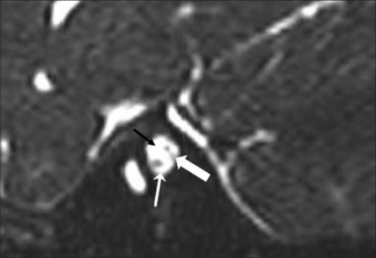
Figure 3.

Parasagittal CISS MR image of left internal auditory canal (IAC) showing absent facial nerve in anterosuperior part of left IAC (black arrow). Vestibular (thick white arrow) and cochlear nerves (thin white arrow) are visualized in posterior and anteroinferior parts of left IAC respectively. (Anterior is to the left of image and posterior is to the right)
