Figure 4.

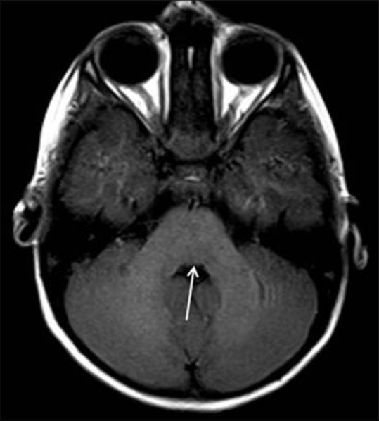
Axial T1W MR image at the level of middle cerebellar peduncles showing flattened floor of fourth (black arrow) ventricle secondary to absence of bilateral facial colliculi

Axial T1W MR image at the level of middle cerebellar peduncles showing flattened floor of fourth (black arrow) ventricle secondary to absence of bilateral facial colliculi