
Figure 4.
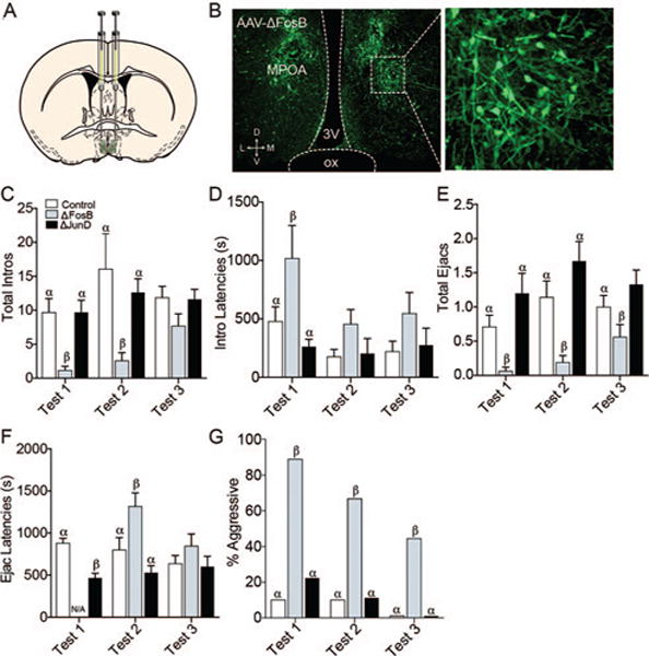
(A) Viral-mediated gene transfer in the MPOA. Diagram illustrates the site of injection at the level of the MPOA. (B) Photomicrograph exemplifies GFP-ir protein as a marker of AAV transfection in the MPOA, 20×. On test one, males receiving ΔFosB had fewer intromissions (C), longer intromission latencies (D), and fewer ejaculations (E) compared to ΔJunD males and GFP controls. Also, ΔJunD males had shorter ejaculation latencies than GFP controls (F). (There were fewer than 3 ΔFosB males ejaculating, so their latencies could not be compared statistically.) On Test 2, ΔFosB males had fewer intromissions (C), ejaculations (E), and longer ejaculation latencies (F) than ΔJunD males and GFP controls. There were no statistical differences in intromission or ejaculation latencies or numbers of intromissions on Test 3. (G) Across all three trials, animals that received ΔFosB were more likely to display female-directed aggression than animals receiving either GFP or ΔJunD. Bars designated with the same letter are not different from each other, but are different from those with the other letter within each test. See the online article for the color version of this figure.