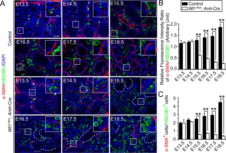
Fig 3. Disruption on the differentiation status of PMC and FLC in Wt1SC-cKO mice that leads to a reduced ratio of PMC:FLC during fetal testis development.
(A) Immunofluorescence analysis of PMC marker α-SMA (TRITC, red fluorescence) and HSD3B1 (FITC, green fluorescence) in cross-sections of control vs. Wt1SC-cKO mouse testes from E13.5 to E18.5. Insets are the corresponding magnified views of the boxed areas. In control testes, the number of α-SMA+ PMCs increased considerably from E13.5 to E18.5 during fetal testis assembly. HSD3B1-stained FLCs were found between testis cords during the development of interstitium. In Wt1SC-cKO testes, deletion of Wt1 in Sertoli cells led to a considerable reduction in PMC number from E15.5 to E18.5. HSD3B1-positive (HSD3B1+) FLCs were found to be differentiated, forming cell clusters from E16.5 to E18.5 in Wt1SC-cKO testes. The α-SMA+ PMCs were found in and around the remnant tubules, and normal testis architecture was not established. (B) The ratio of α-SMA to HSD3B1 relative fluorescence intensity was obtained by measuring the relative fluorescence intensity, which increased in E15.5 to E18.5 in control testes but reduced in Wt1SC-cKO testes, illustrating Wt1 deletion led to changes in the differentiation status of PMC vs. FLC. (C) Consistent with findings shown in (B), the ratio of α-SMA+ PMCs:HSD3B1+ FLCs (α-SMA+ PMCs:HSD3B1+ FLCs) which obtained by scoring these two cell types increased in E15.5 to E18.5 in control testes but reduced in Wt1SC-cKO testes.

