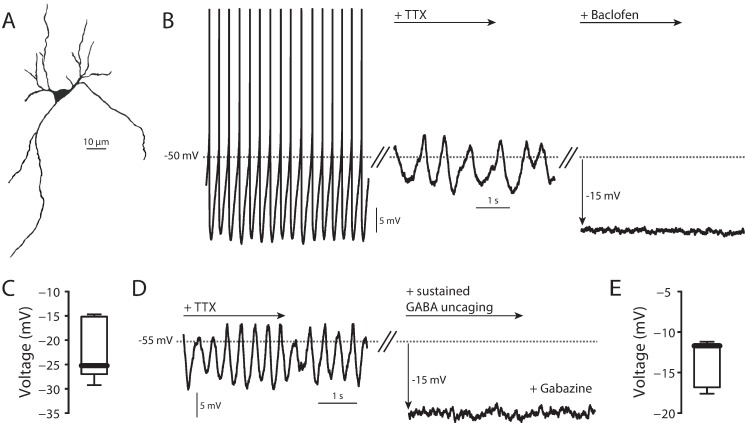
Fig 1. SNc DA neuron physiology.
(A) 2P reconstruction of SNc DA neuron. (B) Left, normal pacemaking of a SNc DA neuron. Middle, TTX (1 μM) application uncovered slow oscillatory potential (SOP). Right, 5 μM baclofen application hyperpolarized the cell. (C) Summary of hyperpolarization due to application of 5 μM baclofen (n = 8, median = -25.24 mV). (D) Sustained uncaging of 5 μM RuBi-GABA in the presence of 25 μM gabazine (to block GABAA receptors) hyperpolarized cells in a manner similar to that seen following baclofen application. (E) Summary of hyperpolarization due to sustained 5 μM RuBi-GABA uncaging (n = 7, median = -11.71 mV).

