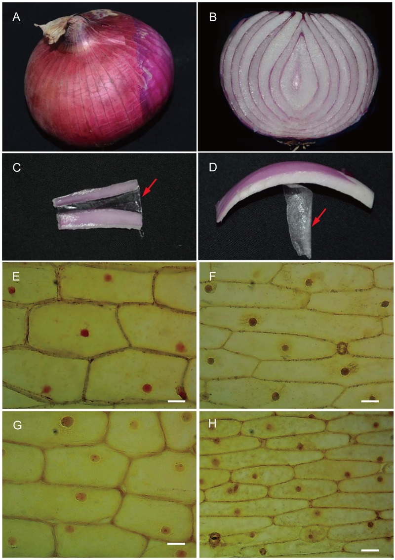
Fig 1. The epidermises of onion scales.
(A) Red onion bulb. B, Longitudinal section of a red onion scale. (C) The lower epidermis (LE) (arrow). (D) The upper epidermis (UE) (arrow). (E-H) Light microscopy of the LE (F and H) and UE (E and G) from a yellow onion scale (E and F) and a red onion scale (G and H). The bar = 2.0 μm.

