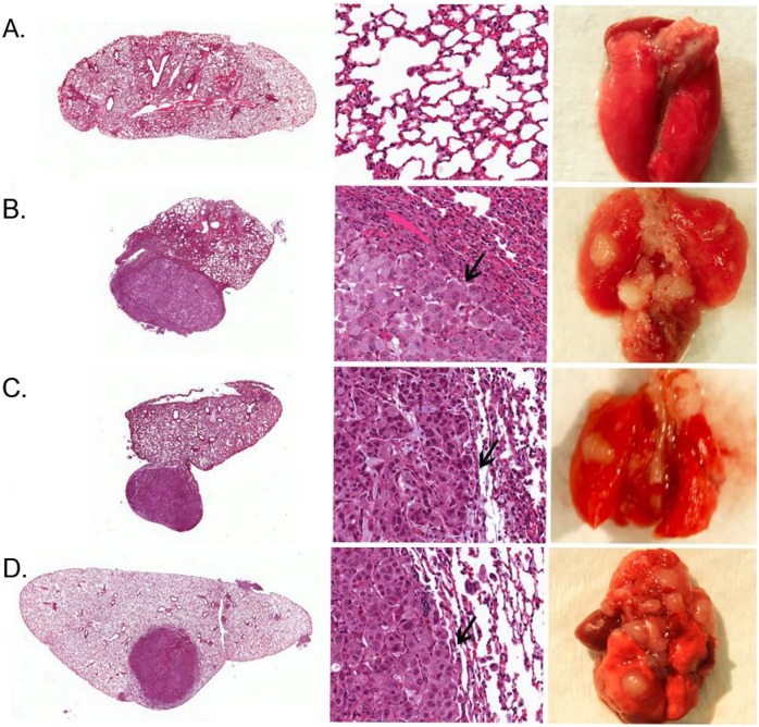
Fig 5. H&E staining of orthotopically-induced lung tumors in athymic Nude-Fox1nu mice.
Tumors were induced by injecting A549-hNIS cells that were genetically modified with either plasmid or lentiviral vectors. Mice were sacrificed at 36 days post cancer cell inoculation. An H&E section of the lung is shown at 4x magnification (left), 60x magnification (middle; arrow marks tumor margin), or (right) gross specimen showing solitary nodules, from the induction of A) Saline control (no tumor). B) A549 lung adenocarcinoma control cell line; no genetic modification. C) A549-pDNA tumor 1. D) A549-pDNA tumor 2. E) A549-LV tumor (n = 3; representative images shown).

