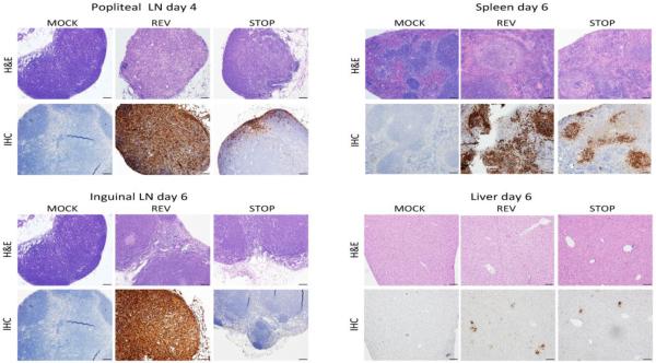
Fig. 6.
Histological examination. Female BALB/c mice (n=3 per group) were infected in the footpad with PBS (mock) or 1000 PFU of C15Rev or C15Stop and sacrificed on days 2, 4, and 6 post-infection. Harvested tissues were fixed with formalin, embedded in paraffin, stained with hematoxylin and eosin (H&E) or with a polyclonal anti-VACV antibody for immunohistochemistry (IHC) and examined via light microscopy. Images representative of the popliteal lymph node on day 4 post-infection and inguinal lymph node, spleen and liver on day 6 post-infection are shown.

