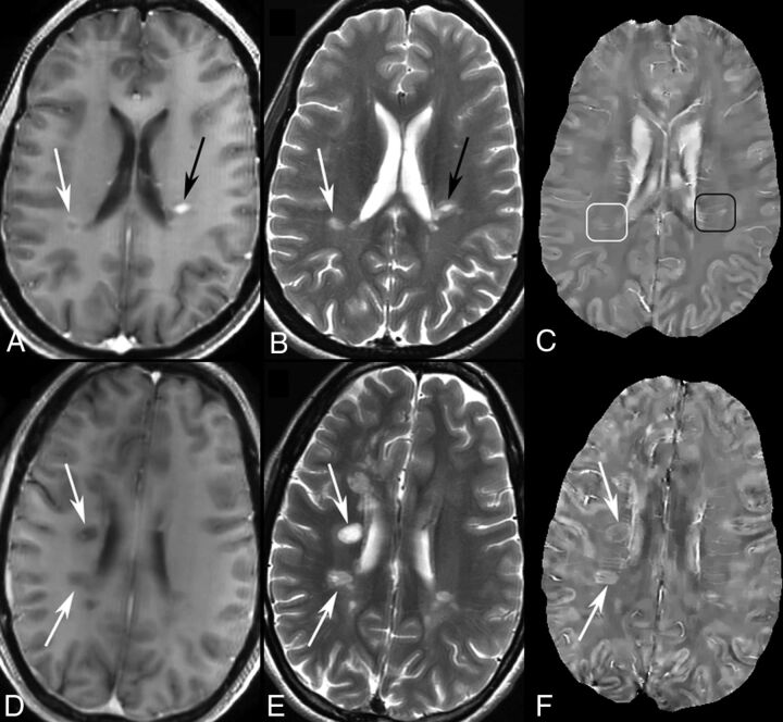
Fig 1.
MR images of enhancing and nonenhancing new MS lesions. T1WI+Gd (A), T2WI (B), and QSM (C) in a 44-year-old woman with relapsing-remitting MS. Two enhancing lesions (A and B, arrows) are found in T1WI+Gd. One is shell-enhancing (A, white arrow) and another is nodular-enhancing (A, black arrow). The shell-enhancing lesion appears slightly QSM-hyperintense (C, white box) and the nodular one appears QSM isointense (C, black box). T1WI+Gd (D), T2WI (E), and QSM (F) in a 35-year-old woman with relapsing-remitting MS. Two new nonenhancing lesions (D and E, arrows) are found in T1WI+Gd and T2WI compared with MR imaging 6 months prior. The 2 lesions both appear QSM-hyperintense with bright rims (F, arrows).

