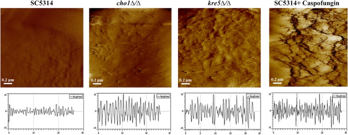
FIG 4.
Topographic 2- by 2-μm images of the cell wall surface of C. albicans wild-type cells with or without caspofungin treatment and cho1Δ/Δ and kre5Δ/Δ mutant cells. Cells were mounted onto gelatin-coated mica and imaged in water in contact mode with cantilevers having spring constants of 0.01 or 0.03 N/m by using a 5500 PicoPlus AFM instrument. PicoPlus AFM software was used to measure surface roughness, which is shown in the plot below each corresponding image. The plot for each strain was the average from measurements made for 30 cells.

