FIG 1.
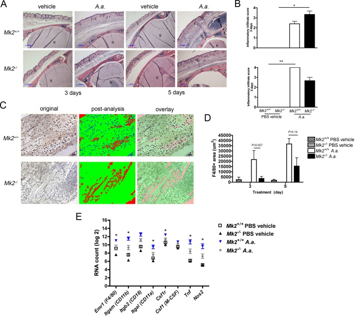
MK2 signaling regulates A. actinomycetemcomitans-induced inflammatory infiltrate. (A) Representative ×40-magnification images of Mk2+/+ and Mk2−/− mouse calvaria, including the epithelium (E), calvarium (C), and brain (B), treated with PBS vehicle or A. actinomycetemcomitans (A.a.) for 3 or 5 days. Blue scale bars, 100 μm. (B) Inflammatory infiltrate scores for Mk2+/+ and Mk2−/− mice treated with PBS vehicle or A. actinomycetemcomitans for 3 days (top) and 5 days (bottom). (C) Representative ×200-magnification images obtained after F4/80 staining of A. actinomycetemcomitans-treated Mk2+/+ and Mk2−/− mice for 3 days and Visiopharm analysis. (D) F4/80-positive (F4/80+) area of Mk2+/+ and Mk2−/− mice treated with PBS vehicle or A. actinomycetemcomitans for 3 and 5 days. (E) Analysis of macrophage marker RNA in mouse calvarial tissue treated for 3 days by use of the NanoString Technologies immunology panel (n = 3 to 5). Data are expressed as means ± SEMs. *, P ≤ 0.05; **, P ≤ 0.01.

