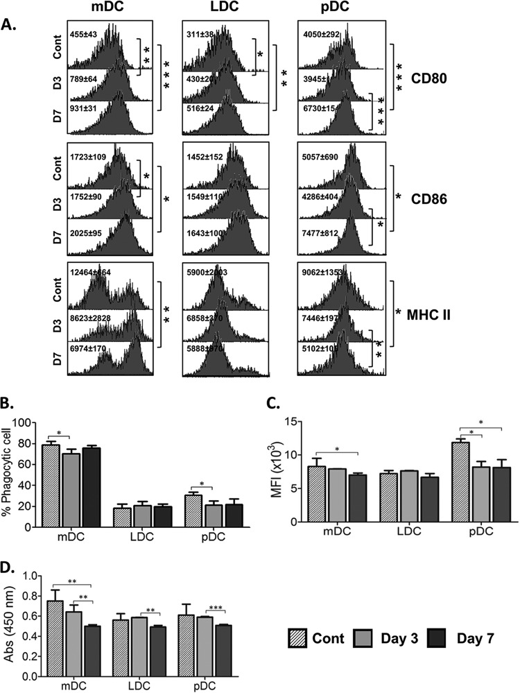
FIG 5.
Assessment of maturation markers and T-cell proliferation capacities of host dendritic cell subsets. (A) Expression of maturation and costimulatory markers was assessed on flow-sorted mDCs, LDCs, and pDCs at day 3 and day 7 post-Bm-L3 infection using flow cytometry. Representative histograms show MFI values of DC subsets at the given time points. (B and C) Antigen uptake (phagocytosis) and antigen presentation capacities of flow-sorted mDCs, LDCs, and pDCs were assessed at day 3 and day 7 post-Bm-L3 infection by incubating the cells with either FITC-dextran or DQ-ovalbumin, respectively, followed by acquisition on a BD FACS Aria flow cytometer, as described in Materials and Methods. Shown are percentages of phagocytic cells (B) and MFIs (C) of DC subsets at the given time points. (D) Mitochondrial activity, as a measure of T-cell proliferation, was measured by XTT assay by coculturing flow-sorted mDCs, LDCs, and pDCs with CD4+ T cells, as described in Materials and Methods. Shown are optical densities at 450 nm (OD450) of DC subsets at the given time points. The data are representative of three independent experiments with at least 5 or 6 animals/group. P values of ≤0.05 (*), ≤0.01 (**), and ≤0.001 (***) were considered significant, highly significant, and very highly significant, respectively.

