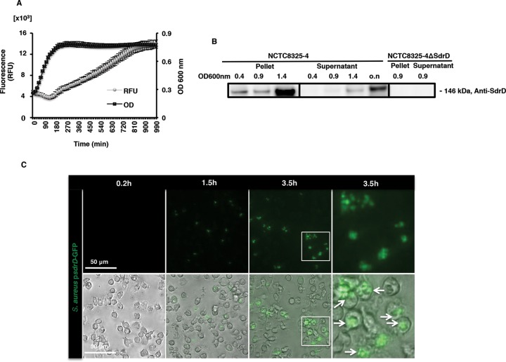
FIG 1.
sdrD promoter activity and SdrD protein expression under different growth conditions. (A) Promoter activity of sdrD during growth in TSB using NCTC8325-4 harboring the sdrD-GFP reporter construct (S. aureus subsp. aureus psdrD-GFP). Data represent the means ± SEMs from an individual experiment. The experiments were performed twice in triplicate. RFU, relative fluorescence units. (B) Immunoblotting of the bacterial lysates and the culture cell-free supernatant of NCTC8325-4 and its isogenic mutant, NCTC8325-4 ΔsdrD, using anti-SdrD antibody on the bacterial lysates and the culture cell-free supernatant. A representative Western blot is shown. (C) Promoter activity of sdrD evaluated by fluorescence microscopy using NCTC8325-4 harboring psdrD-GFP in the presence of freshly isolated human neutrophils. (Top) Live imaging was performed after 0.2, 1.5, and 3.5 h using fluorescence microscopy; (bottom) bright-field and GFP merged images. The white boxes in the first column labeled 3.5 h are enlarged in the second column labeled 3.5 h. The experiment was performed at an MOI of 20. Arrows, S. aureus subsp. aureus psdrD-GFP. The sizes of the scale bars are indicated.

