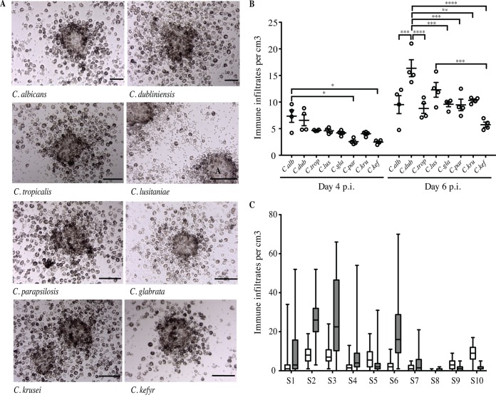
FIG 1.
In vitro immune infiltrates induced by Candida spp. after infection of human immune cells from immunocompetent subjects. (A) Representative immune infiltrates observed under light microscopy 6 days after challenge of human mononuclear and polymorphonuclear peripheral blood cells with living yeasts from annotated Candida species (MOI, phagocyte-to-yeast ratio of 2,000:1). Bars represent 50 μm. No formation of immune infiltrates was observed in uninfected conditions for up to 6 days postinfection (p.i.). (B) Number of immune infiltrates per cubic centimeter on days 4 and 6 postinfection by the eight Candida species. Each dot represents the mean of the number of structures per cubic centimeter for one clinical isolate and for all studied subjects. Lines indicate SEM (*, P between 0.045 and 0.0360; **, P = 0.0017; ***, P between 0.0004 and 0.0002; ****, P < 0.0001; α = 0.05). C.alb, C. albicans; C.dub, C. dubliniensis; C.trop, C. tropicalis; C.lus, C. lusitaniae; C.gla, C. glabrata; C. par, C. parapsilosis; C.kru, C. krusei; C.kef, C. kefyr. (C) Box plots depict median, minimum, and maximum immune infiltrate numbers for each donor subject (S) on days 4 (white boxes) and 6 (gray boxes) postinfection (n = 80).

