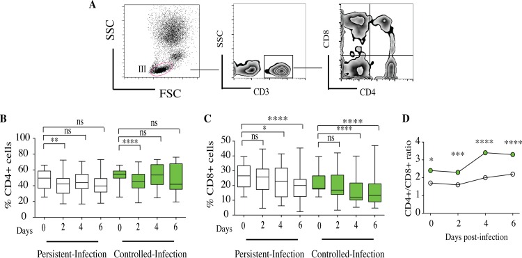
FIG 8.
Characterization of CD4+ and CD8+ T cells within Candida granuloma-like structures over time. Peripheral blood mononuclear and polymorphonuclear cells from 10 subjects were infected with 32 Candida clinical isolates for up to 6 days. The granuloma-like structures were collected from coculture plates at different time points and stained with a cocktail of fluorescence-conjugated antibodies specific to CD3, CD4, and CD8 lymphocytes. (A) Representative flow cytometry analysis showing CD4+ and CD8+ cells over time after gating on CD3+ lymphocytes. (B and C) The proportions of CD4+ and CD8+ cells within granuloma-like structures are expressed as percentages of the total CD3+ compartment. Box plots depict median, minimum, and maximum percentages of CD4+ and CD8+ cells over time according to infection status. (D) Dynamics of CD4+/CD8+ cell ratio over time between persistent-infection and controlled-infection granuloma-like structures. *, P < 0.05; **, P < 0.001; ***, P < 0.0001; ****, P < 0.00001 (by one-way ANOVA with Tukey's multiple-comparison test) (n = 320).

