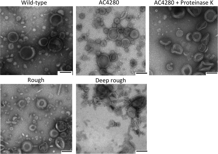
FIG 3.
Transmission electron microscopy examination of outer membrane vesicle morphology. OMVs purified from the strains indicated were diluted to a concentration of 1 μg/μl in PBS. Grids were floated in OMV solution for 1 min, washed with 2% acidic uranyl acetate, and blotted dry before visualization under TEM. Scale bars are 100 nm.

