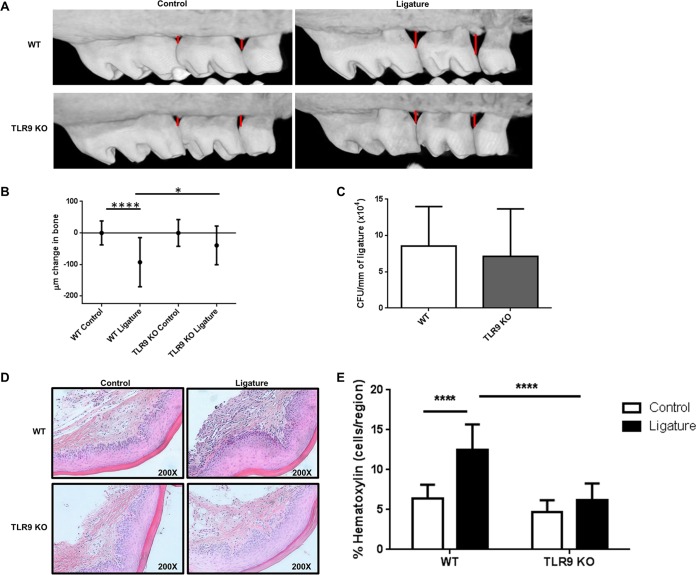
FIG 1.
TLR9 deficiency prevents ligature-induced periodontal bone loss in vivo. On day 7 following ligature placement, WT and TLR9−/− mice were sacrificed, and entire heads were processed for micro-CT imaging. (A) Representative micro-CT images of maxillae from each group of animals. (B) Amounts of bone change in WT and TLR9−/− mice. Negative values represent bone loss in ligated mice relative to the results for the control (unligated) mice. Ten to 18 mice were analyzed. Whiskers show standard deviations. (C) CFU enumeration of periodontal microflora recovered from the ligatures of WT and TLR9−/− mice. The numbers were determined by extracting bacteria from the ligatures and plating serial dilutions of bacterial suspensions onto blood agar plates for anaerobic growth. Ligatures from six to seven mice were included. The average results and standard deviations are shown. (D and E) Gingival tissues were harvested, sectioned, and processed for H&E staining. Representative images of gingival tissue sections from 4 WT and 4 TLR9−/− mice (D) and quantification of inflammatory cells in the tissue sections (E) are shown for ligated and control tissue sections. Tissue specimens from four mice were analyzed in triplicates to sextuplicates in a minimum of 2 independent experiments, and average results and standard deviations are shown. *, P < 0.05; ****, P < 0.0001.

