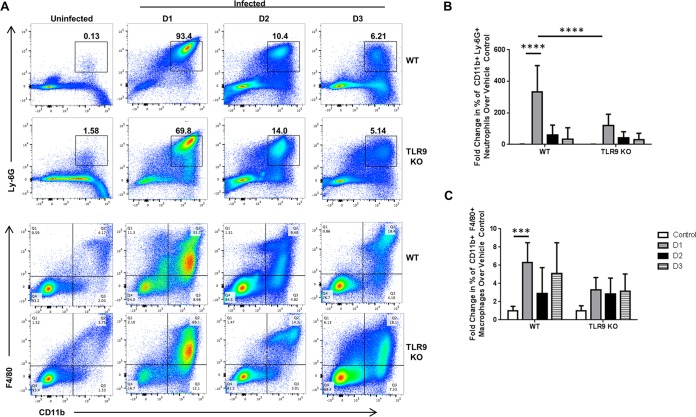
FIG 2.
Decreased inflammatory cell infiltrate in the peritoneal cavities of TLR9−/− mice following Porphyromonas gingivalis challenge. WT and TLR9−/− mice were intraperitoneally injected with 1 × 10⁸ CFU of Porphyromonas gingivalis, and the peritoneal exudate cells were harvested and stained with anti-TCR-β, anti-B220, anti-CD11b, anti-Ly-6G, and anti-F4/80 antibodies. (A) Representative flow cytometry dot plots: the numbers in the panels are representative of the percentage of cells in the respective quadrant or gate. (B and C) Quantification of the percentages of neutrophils (B) and macrophages (C) from the peritoneal lavage exudate samples from mice sacrificed at the indicated time points. The average results and standard deviations are shown. Five to 13 mice were analyzed in a minimum of two independent experiments. ***, P < 0.001; ****, P < 0.0001.

