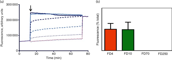
Fig. 3.
VP4N-induced membrane permeability is enhanced by myristoylation. (a) CF release assays showing membrane permeability induced by myristoylated VP4N-45mer at 1 µM (solid line, dark blue), 0.1 µM (dashed line, dark blue) or 0.01 µM (dotted line, dark blue) or unmyristoylated VP4N-45mer at 1 µM (solid line, light blue), 0.1 µM (dashed line, light blue) or 0.01 µM (dotted line, dark blue). Data shown are representative of three independent experiments. The black arrow indicates time of addition of sample at the start of the assay. (b) Release from liposomes of FITC-labelled dextrans of 4 kDa (FD4), 10 kDa (FD10), 70 kDa (FD70) or 250 kDa (FD250) by 0.1 µM myristoylated VP4N-45mer. Data are presented as percentage of total release observed by lysis of liposomes by addition of detergent. Error bars represent standard error of the mean of values from three independent experiments.

