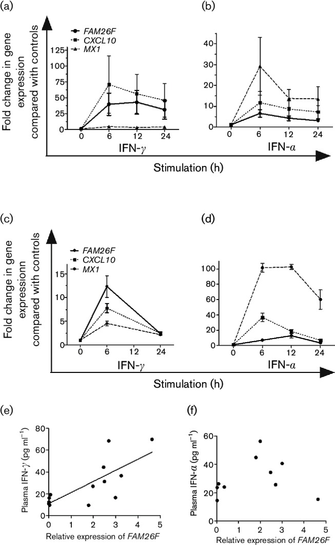
Fig. 3.

FAM26F expression is linked to IFN-γ pathway. IFN-γ was added to PBMCs from (a) eight LTNPs without and (c) two naive macaques with prior low-dose ConA activation (10 ng ml−1 overnight). IFN-α2 was added to PBMCs from (b) eight LTNPs without and (d) three naive macaques with prior low-dose ConA activation. At the time points indicated, RNA was extracted and fold differences in gene expression of FAM26F, CXCL10 and MX1 compared with controls not treated with interferons were determined. Mean and standard error of the mean are shown. (e) Plasma IFN-γ and (f) plasma IFN-α were quantified by ELISA while relative expression of FAM26F and CXCL10 was quantified by RT-PCR in PBMCs. FAM26F expression correlated with plasma IFN-γ (P=0.0031, Pearson r=0.7508; n=13) but not with plasma IFN-α (n=10).
