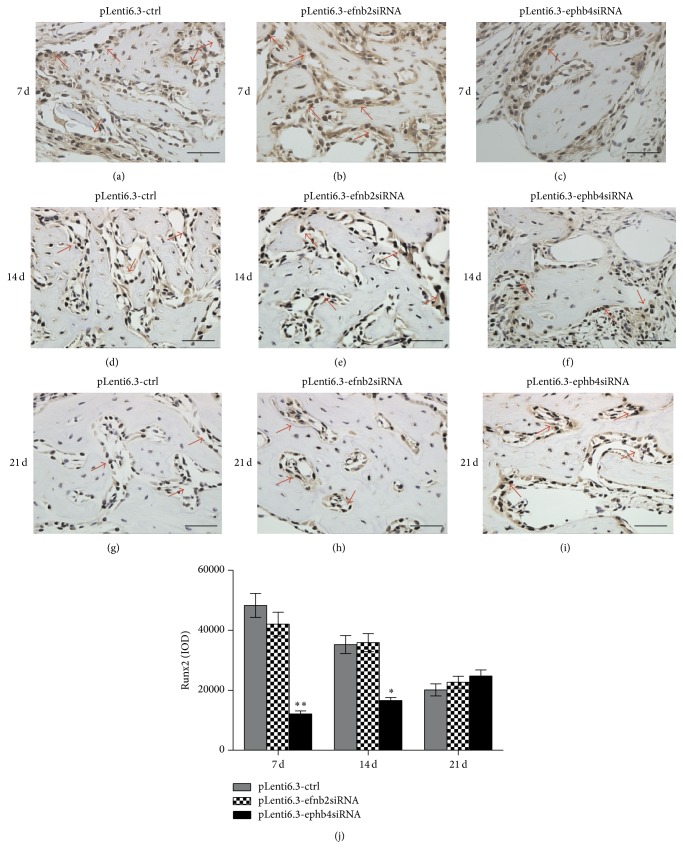
Figure 7.
Immunohistochemistry staining for Runx2. (a–i) At 7 days (a–c), 14 days (d–f), and 21 days (g–i) after surgery, different numbers of Runx2-positive cells with brown-stained nuclei could be found surrounding the trabecular bone in the newly formed bone area in the pLenti6.3-ctrl group (a, d, g), the pLenti6.3-efnb2siRNA group (b, e, h), and the pLenti6.3-ephb4siRNA group (c, f, i). (j) Quantitative analysis via IOD measurements using the Image-Pro Plus 6.0 software. Arrow, positive Runx2 staining; scale bars, 50 μm; ∗ p < 0.05, ∗∗ p < 0.001 versus pLenti6.3-ctrl group.

