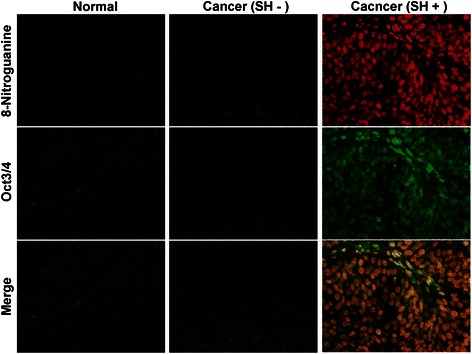
Fig. 9.

Formation of 8-nitroguanine and expression of Oct3/4 in bladder tissues. The formation of 8-nitroguanine (red) and the expression of Oct3/4 (green) were assessed by double immunofluorescence staining [21]. In the merged image, co-localization of 8-nitroguanine and Oct3/4 is indicated in yellow. Biopsy and surgical specimens were obtained from normal subjects and patients with SH-induced cystitis and bladder cancer. Normal tissues and urinary bladder cancer tissues without SH-infection were obtained from a commercial urinary bladder tissue array (Biomax.us, USA)
