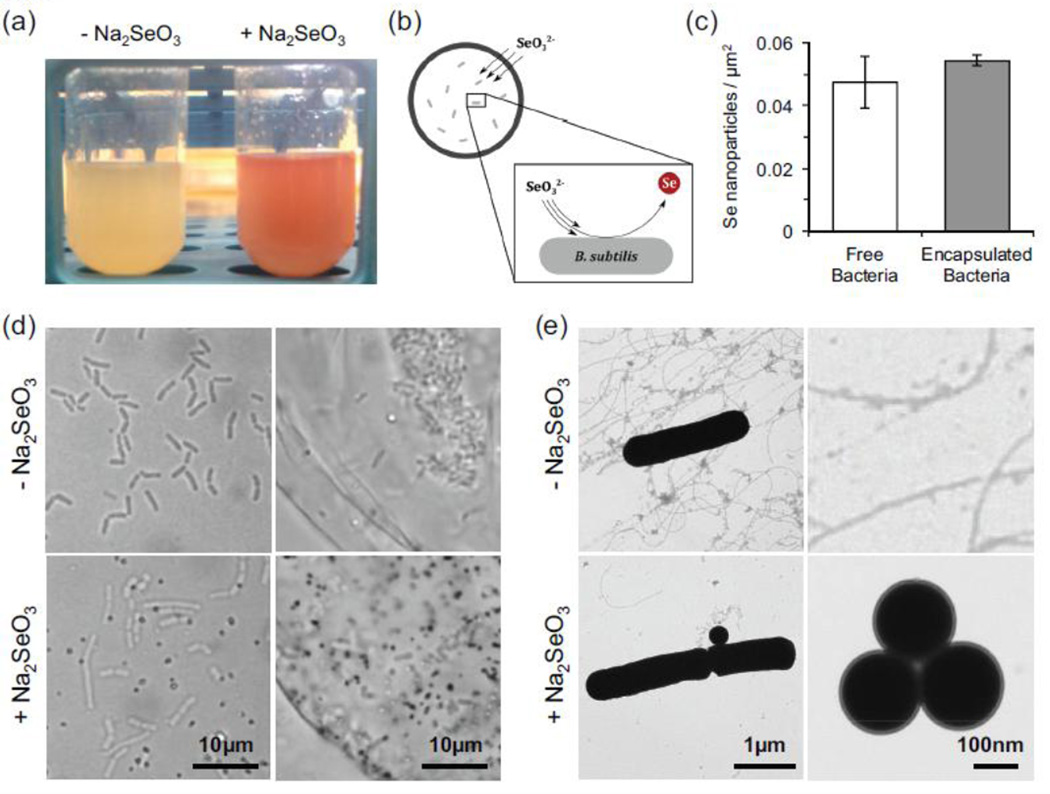
Fig. 5.
Conversion of soluble selenite (Na2SeO3) to elemental selenium (Se) by free and encapsulated bacteria. (a) Eight hours after sodium selenite addition, B. subtilis solutions turn visibly red, indicative elemental selenium nanoparticle formation. (b) Proposed model of selenium remediation. (c) Quantification of selenium nanoparticle synthesis by free and encapsulated bacteria, as measured by optical microscopy. (d) Widefield images of encapsulated bacteria in the presence and absence of Na2SeO3. The addition of Na2SeO3 to the supernatant results in formation of selenium nanoparticles (dark dots) by free and encapsulated B. subtilis. (e) Transmission electron microscopy images of nanoparticle formation by B. subtilis.

