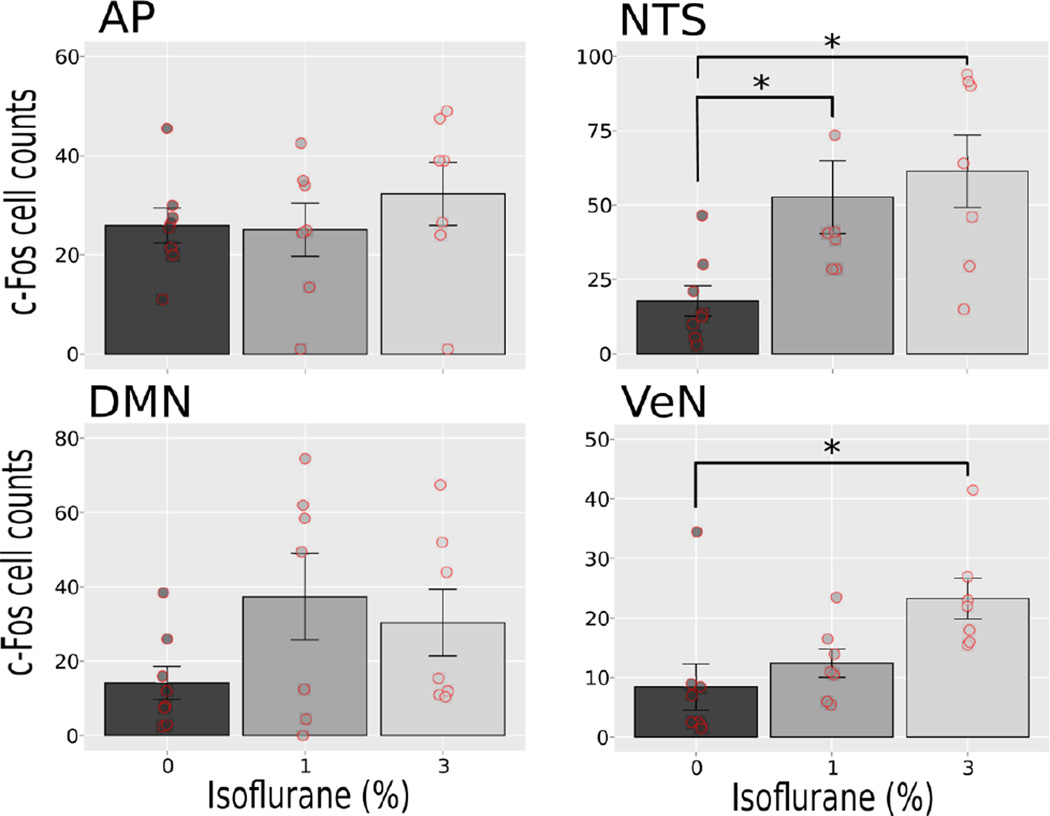
Fig. 5.
Study 2: Quantification of c-Fos-positive cells at 0, 1 and 3% isoflurane exposures. Brain areas include the area postrema (AP), nucleus of the solitary tract (NTS), dorsal motor nucleus (DMN), and the vestibular nuclei (VeN). Values are means ± SEM, with scatter plots of the raw values. * = p < 0.05, LSD-tests.

