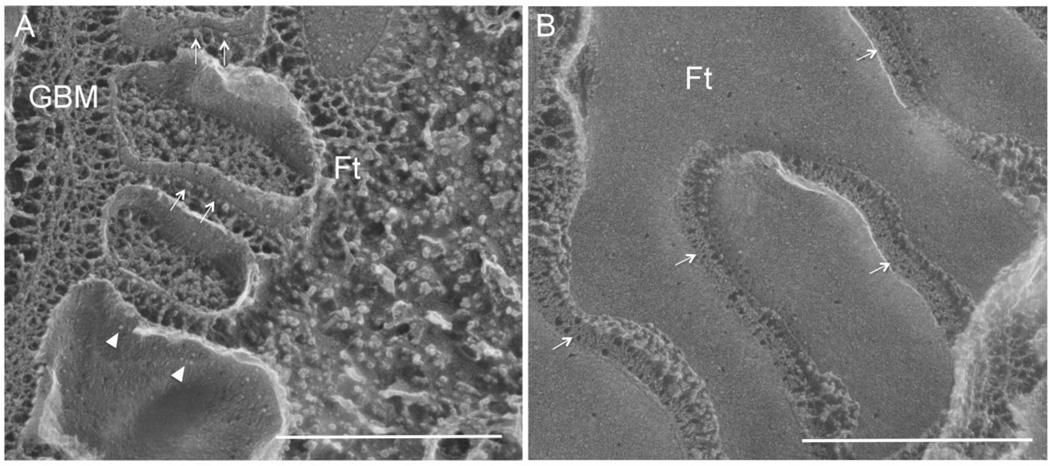
Figure 3.
Freeze fracture electron micrographs showing the replica of fractured podocytes from both the perpendicular view (A) and the parallel view (B) relative to the glomerular basement membrane. In A, arrow indicates the slit diaphragm as protein particles anchored on the E-face of the fractured membrane and arrowhead indicates the slit diaphragm protein particles on the P-face of the fractured membrane. In B, arrow indicates the slit diaphragm as zipper-like structure. GBM: glomerular basement membrane; Ft: foot process. Bar: 500nm. Adapted from reference (28).

