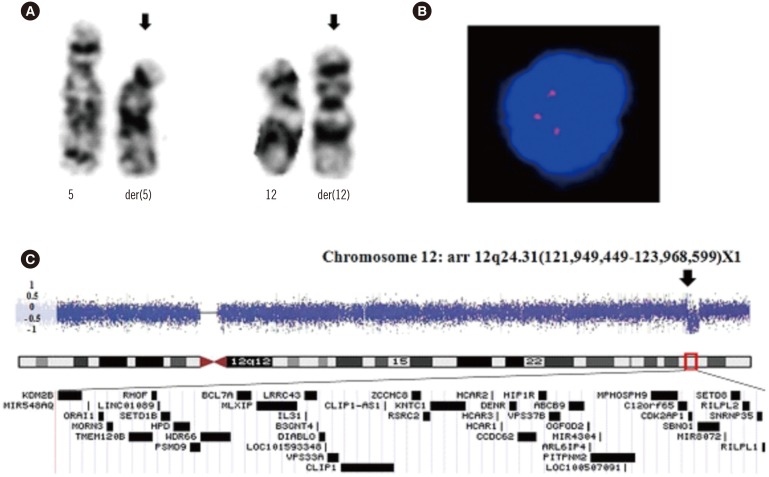
Fig. 1. Genetic analysis of CML-AP BM samples. (A) Translocation site in G-banded karyotyping of the 46,XY,t(5;12)(p13;p13) (arrows). (B) Interphase FISH confirming three signals of the AID probe (red), resulting from AID rearrangement at the 12p13 locus. (4,6-diamidino-2-phenylindole stain, ×1,000). (C) Chromosomal microarray revealing copy number loss in the chromosome 12q24.31 region (arrow). Blue dots with a log2 transformed value of -1 represent a 1:2 copy number ratio to the reference genomic DNA, indicating a heterozygous deletion. The expansion view of the 12q24 region reveals a 1.9-Mb heterozygous interstitial deletion in chromosome 12 (121,949,449-123,968,599; hg19) that includes the BCL7A gene.
Abbreviations: AP, accelerated phase; BM, bone marrow; AID, activation-induced cytidine deaminase; BCL7A, B-cell CLL/lymphoma 7A.

