. 2016 Dec 23;19(4):429–437. doi: 10.4048/jbc.2016.19.4.429

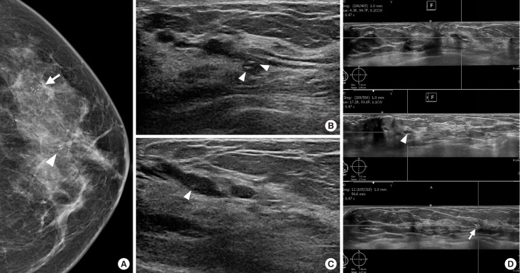
Figure 5. A 43-year-old woman with ductal carcinoma in situ in the left breast. (A) Craniocaudal view of mammogram shows segmental fine pleomorphic microcalcifications in central (arrowhead) and peripheral outer breast (arrow). (B, C) Handheld ultrasound (US) shows an intraductal hypoechoic mass with microcalcifications (arrowheads), which matched with central microcalcifications on mammogram. (D) Automated whole breast US shows segmental ductal dilatation with intraductal microcalcifications (arrowhead), which matched with central microcalcifications on mammogram. Grouped microcalcifications (arrow) in peripheral breast are found, which matched with peripheral microcalcifications on mammogram.

Figure 5