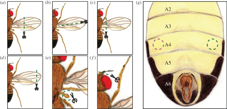
Figure 1.
Schematic of the different injuries made to the flies. (a) This was the standard procedure, where the distal two-thirds from both wings were removed. (b) Longitudinal cut. Half of the wing was removed. This was applied to both wings in experiments of figure 4a,b. (c) Whole wing cut. This was used in figure 4c,d to remove only one wing (the side was randomly selected), and in figure 4e,f to remove both wings. (d) End of the wing cut. Around 20% of each wing was removed. It was used in figure 4e,f. (e) Haltere removal. Both halteres were removed and the effect on photopreference is presented in figure 4g,h. (f) Antennal damage. The third segment of both antennae was cut. This treatment was used for experiments in figure 4i,j. (g) Abdominal injury. Flies were stabbed on one side of the ventral fourth abdominal segment (the side was randomly selected). The results of the effect of this injury in phototaxis are depicted in figure 4k,l.

