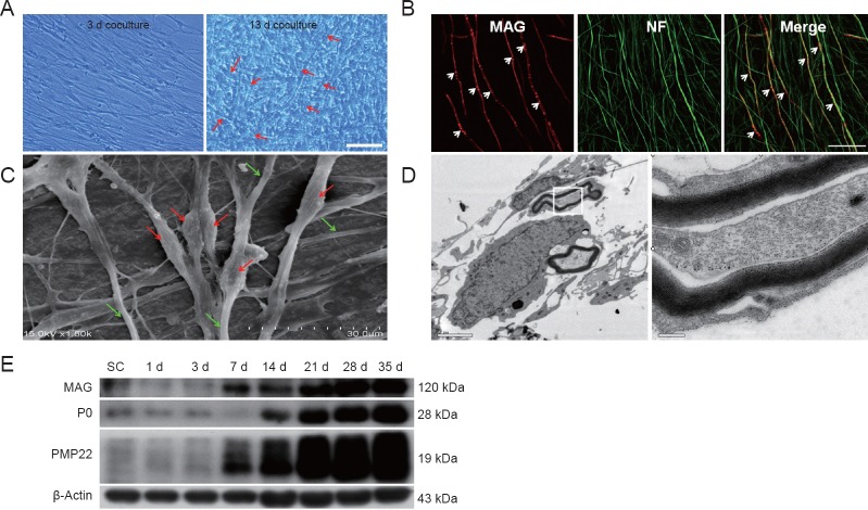
Figure 2.
Establishing a myelination model of DRG neurons co-cultured with Schwann cells.
(A) Phase-contrast microscopy at 3 and 13 days after the establishment of a DRG neuron and Schwann cell co-culture. Arrows indicate myelin sheath-like structures. Scale bar: 50 μm. (B) Double-staining with anti-MAG (red) and anti-NF (green) to examine myelination. Arrows indicate segregation of MAG staining in the neurites of DRG neurons. Scale bar: 50 μm. (C) Scanning electron microscopy shows Schwann cells surrounding DRG neuron neurites and the formation of myelin segments. The diameters of the myelinated axons are thicker (red arrows) than the neuritis of DRG neurons (green arrows). Scale bar: 30 μm. (D) Transmission electron microscopy also shows the typical compact myelin structure. Right: High-magnification image from inset box. Scale bar: 2 μm on the left and 0.2 μm on the right. (E) Western blot images of expression of myelin proteins MAG, P0, and PMP22 at 1, 3, 7, 14, 21, 28 and 35 days after co-culture. β-Actin served as a protein loading control. Compared with purified Schwann cells alone, the DRG neuron and Schwann cell co-cultures induce a significant increase in all myelin proteins. DRG: Dorsal root ganglion; d: days.

